 Anatomy
Dogs have 13 ribs that form the supportive structure (rib cage) of the chest cavity. The ribs attach onto the breastbone and the backbones. The vital structures such as the heart and the lungs are protected by the rib cage. The muscles of the chest pull on the ribs during inspiration to increase the size of the chest cavity to pull air into the lungs. As the muscles relax, the rib chest cavity size gets smaller and air is breathed out.
Types of rib tumors
The most common type of rib tumor is osteosarcoma. This is a very malignant tumor and has a rapid spread rate (metastatic). The second most common tumor is chondrosaroma. This tumor has a much lower metastatic potential. Other common tumors that affect the ribs include fibrosarcoma and hemangiosarcoma.
Signs
 Breeds most commonly to develop rib tumors included Golden Retrievers, mix breeds, Labrador Retrievers, Bassett Hounds, Dobermans, Australian Shepherds, British Bulldog, German Short-haired Pointer, Rough coated Collie, Irish Setter, Giant Pyrenees, Rottweiler, Giant Schnauzer, Shar Pei, and Springer Spaniel. The age of dogs that develop rib tumors tends to be a bimodal distribution with affected dogs being either young (2 to 4.5 years) or older (7 to 9 years). The most common sign of a rib tumor is a mass that is visibly or palpably present. Lameness of the forelimb may be present if the tumor is located within one of the first four ribs, is compressing the nerves to the limb, is causing mechanical interference with movement of the limb or is invading into the muscles of the forelimb. Labored breathing may be noted if the tumor is very large and is causing collapse of the lung. If the tumor has metastasized to the inside of the chest cavity, fluid may build up in the chest and compress the lungs, thus cause labored breathing.
 Diagnosis
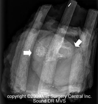
The diagnosis of a rib tumor is commonly made on x-rays. Typically, the tumor will cause a portion of the rib bone to be dissolved away and/or new bone to be produced within the region of the tumor or both (photo right). Chest x-rays (three views) should be made to rule out visible spread of the tumor to the lungs. A CT scan may be recommended, as this test provides a better evaluation of the extent of the disease and is more sensitive to detect evidence of spread of the tumor into the lungs. A fine needle biopsy may be helpful to diagnose a rib tumor, but frequently will not ascertain the specific type of tumor that is present. Usually the entire tumor is submitted for biopsy after surgery is preformed to establish a final diagnosis. If available, a whole body nuclear bone scan is recommended prior to surgery, as roughly 16%, of the rib osteosarcomas will have spread to other bones at the time of diagnosis. In preparation for surgery, preoperative blood work including a complete blood count, chemistry profile and urine testing are recommended to ensure that your pet is adequately healthy to under go anesthesia and surgery.
The day of surgery
Please make sure that your companion has been fasted prior to surgery and that the prescribed dose of Pepcid AC has been administered. The surgeon will contact you after the surgery to give you a progress report on your companion after the surgery. Our anesthesia and surgical team will prescribe a pain management program, both during and after surgery that will keep your companion comfortable. This will include a combination of general anesthesia, injectable analgesics, local anesthetics, oral analgesics and anti-inflammatory medication.
Treatment
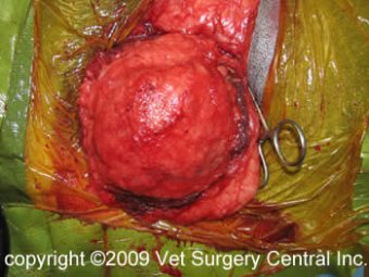
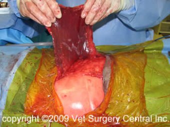
Surgery is the recommended treatment option for rib tumors. In addition to the rib(s) that are affected by the tumor, one unaffected rib in front and behind the tumor should be removed (photo right). If the tumor is adherent or invading into a lung lobe or the sac surrounding the heart, this tissue will also be removed. In dogs, this is generally tolerated very well. In some cases, a plastic mesh patch will be used to repair the defect in the rib cage. In most cases, a muscle (latissimus dorsi or external abdominal oblique muscle) will be used to repair the defect in the rib cage (photos below left and below right). A drainage tube is placed within the cast cavity and another drainage tube may be placed beneath the skin and muscle flap.
YOU MIGHT ALSO LIKE
Share this Post
latest post
-
 Full grown Shar Pei dog February 17, 2017
 -
 Bear Shar Pei June 1, 2018
 -
 Shar Pei, Rottweiler March 3, 2017
 -
 Shar Pei Labrador Retriever mix June 10, 2016
 -
 Shar Pei growth chart April 15, 2016
 -
 Best dogs food for Shar Pei February 3, 2016
 -
 Toy Shar PEI for sale April 27, 2018
 -
 Chow Chow and Shar Pei mix April 14, 2017
 -
 Shar PEI harness March 8, 2016